Muscles Of The Torso / a view of the most superficial posterior muscles of the ... : Terms in this set (68) rectus femoris.. There is a printable worksheet available for download here so you can take the quiz with pen and paper. Each rib attaches to the spine at the back of the body and then. The coracobrachialis and pectoralis major muscles connect the humerus anteriorly to the scapula and ribs, flexing and adducting the arm toward the front of the body when you reach forward to grab an object. Aponeurosis of the abdominal external oblique muscle. The muscles of the trunk include those that move the vertebral column, the muscles that form the thoracic and abdominal walls, and those that cover the pelvic outlet.
Back muscle anatomy chart 12 photos of the back muscle anatomy chart back muscle anatomy chart, lower back muscle anatomy chart, human muscles, back muscle anatomy chart, lower back muscle anatomy chart Muscles of the lower body. Human muscles · august 21, 2016. Identify the movement and function of the intrinsic skeletal muscles of the back and neck, and the skeletal muscles of the abdominal wall and thorax. Working as a team, these muscles contract to flex, laterally bend, and rotate the torso.
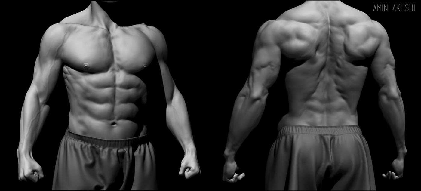
Male Torso on Behance from mir-s3-cdn-cf.behance.net If these muscles have spasms, you feel painful, tightening pressure. The chest comprises of 12 rib bones on each side of the body. You use this muscle when you stand up, walk, run, and climb stairs in fact—whenever you straighten or extend your legs. Muscles of the lower body. Ibs often leads to pain in the colon, which is located on the left side of the abdomen, that can sometimes be accompanied by chronic spasming of the abdominal muscles. Linda nylind for the guardian. You might hear a doctor call it dysesthesia, which means a sensation that isn't normal. Hamstrings (semi membranosus, semiteninosus, biceps femoris), gastrocnemius, popliteus extensors:
The gluteus maximus is the largest muscle in the body.
The main movements of the knee are flexion and extension.for lateral knee pain, look at the vastus lateralis. Neck flexion refers to the motion used to touch the chin to the chest. Muscles the major muscle in the chest is the pectoralis major. These muscles attach the upper limb to the axial skeleton of the trunk and support the thoracic cage. The muscles of the trunk include those that move the vertebral column, the muscles that form the thoracic and abdominal walls, and those that cover the pelvic outlet. Mississippi gulf coast community college. Again, this view of the muscles of the torso seen from the side is obviously informed by dissection, even though these are superficial muscles which show clearly under the surface of the skin. This is one of the internal rotator muscles that attach the humerus and internally rotate the arm. The dominant muscle in the upper chest is the pectoralis major. The muscles of the human body can be categorized into a number of groups which include muscles relating to the head and neck, muscles of the torso or trunk, muscles of the upper limbs, and muscles of the lower limbs. The muscles of the vertebral column, thorax, and abdominal wall extend, flex, and stabilize different parts of the body's trunk. This list may not reflect recent changes. Terms in this set (68) rectus femoris.
The intercostal muscles form the chest wall and function in respiration. Back muscle anatomy chart 12 photos of the back muscle anatomy chart back muscle anatomy chart, lower back muscle anatomy chart, human muscles, back muscle anatomy chart, lower back muscle anatomy chart The dominant muscle in the upper chest is the pectoralis major. The main movements of the knee are flexion and extension.for lateral knee pain, look at the vastus lateralis. A side strain is a condition that is commonly seen in cricket fast bowlers and is characterized by a tear in one of the side abdominal muscles (known as the internal oblique) where it attaches to the lower ribs.
Part 7 - Muscles of the human body - YouTube from i1.ytimg.com The dominant muscle in the upper chest is the pectoralis major. Muscles the major muscle in the chest is the pectoralis major. Identify the movement and function of the intrinsic skeletal muscles of the back and neck, and the skeletal muscles of the abdominal wall and thorax. The main movements of the knee are flexion and extension.for lateral knee pain, look at the vastus lateralis. You need to get 100% to score the 16 points available. Muscle charts of the human body for your reference value these charts show the major superficial and deep muscles of the human body. The oblique muscles down your side are an important area to focus on if you want. Aponeurosis of the abdominal external oblique muscle.
This is an online quiz called torso muscles.
Identify the movement and function of the intrinsic skeletal muscles of the back and neck, and the skeletal muscles of the abdominal wall and thorax. Mississippi gulf coast community college. Again, this view of the muscles of the torso seen from the side is obviously informed by dissection, even though these are superficial muscles which show clearly under the surface of the skin. Related posts of muscles on the side of your torso back muscle anatomy chart. Each rib attaches to the spine at the back of the body and then. Superficial and deep anterior muscles of upper body The erector spinae muscles (iliocostalis, longissimus, and spinalis) are large, deep muscles that extend the length of the back. The oblique muscles down your side are an important area to focus on if you want. A chest muscle that pulls the arm in towards the body. If these muscles have spasms, you feel painful, tightening pressure. Identify what the arrow is pointing to. Pages in category muscles of the torso the following 48 pages are in this category, out of 48 total. Watch the whole lecture (all 8 videos) by goin.
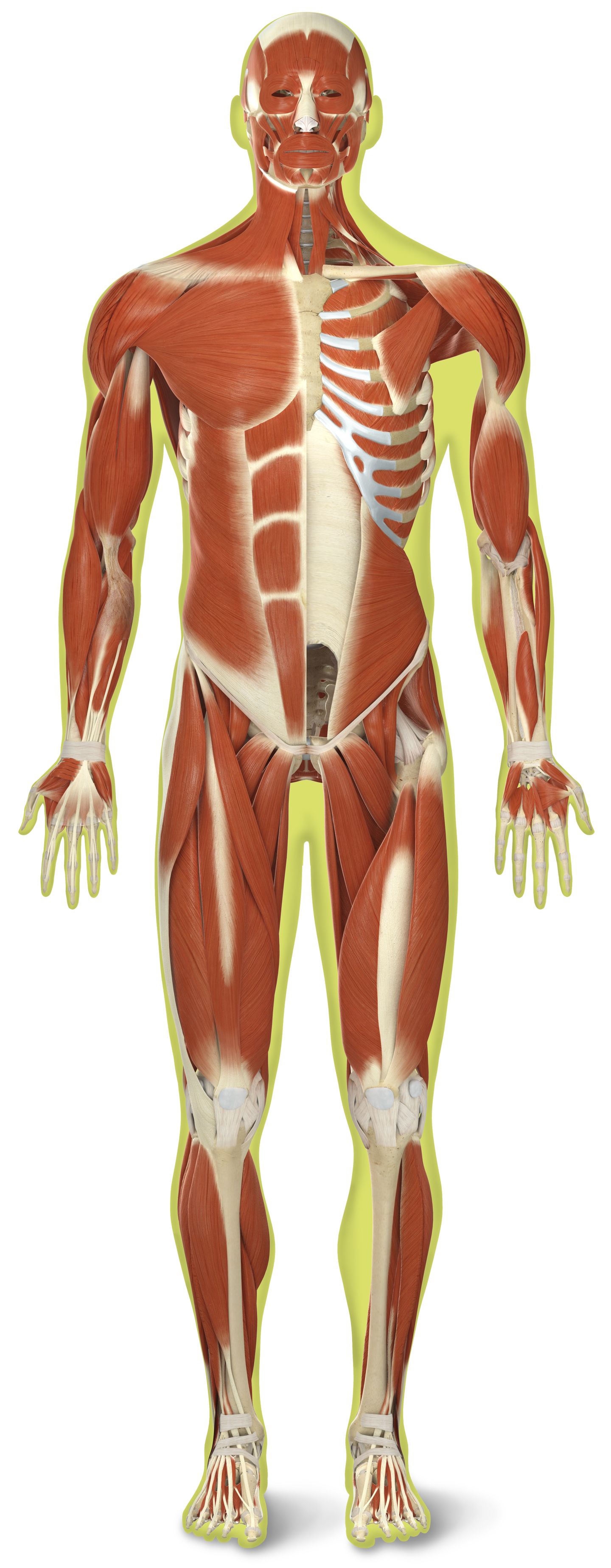
The main movements of the knee are flexion and extension.for lateral knee pain, look at the vastus lateralis. Superficial and deep anterior muscles of upper body The muscles of the vertebral column, thorax, and abdominal wall extend, flex, and stabilize different parts of the body's trunk. The muscles of the trunk include those that move the vertebral column, the muscles that form the thoracic and abdominal walls, and those that cover the pelvic outlet. Hamstrings (semi membranosus, semiteninosus, biceps femoris), gastrocnemius, popliteus extensors:
Skeletal Muscles | Skeletal Muscle Definition | DK Find Out from res.cloudinary.com The dominant muscle in the upper chest is the pectoralis major. The erector spinae muscles (iliocostalis, longissimus, and spinalis) are large, deep muscles that extend the length of the back. The hug is a type of nerve pain. Superficial and deep anterior muscles of upper body The coracobrachialis and pectoralis major muscles connect the humerus anteriorly to the scapula and ribs, flexing and adducting the arm toward the front of the body when you reach forward to grab an object. It is a complex job to balance the body on two feet and walk upright. Pages in category muscles of the torso the following 48 pages are in this category, out of 48 total. Each rib attaches to the spine at the back of the body and then.
Each rib attaches to the spine at the back of the body and then.
You use this muscle when you stand up, walk, run, and climb stairs in fact—whenever you straighten or extend your legs. Why is my entire torso cramping? Again, this view of the muscles of the torso seen from the side is obviously informed by dissection, even though these are superficial muscles which show clearly under the surface of the skin. Watch the whole lecture (all 8 videos) by goin. Superficial and deep anterior muscles of upper body In the lower sketch leonardo shows some of the deeper layer of muscles as they work under the. The intercostal muscles form the chest wall and function in respiration. It runs from the back of the pelvis to the upper part of the femur. Identify the movement and function of the intrinsic skeletal muscles of the back and neck, and the skeletal muscles of the abdominal wall and thorax. These muscles attach the upper limb to the axial skeleton of the trunk and support the thoracic cage. The muscles of the trunk include those that move the vertebral column, the muscles that form the thoracic and abdominal walls, and those that cover the pelvic outlet. You might hear a doctor call it dysesthesia, which means a sensation that isn't normal. Identify what the arrow is pointing to.